Wax-filled stomatal antechambers are excellent antitranspirants.


Epicuticular wax in the stomatal antechamber of sitka spruce and its effect on the diffusion of water vapour and carbon dioxide

by Jeffree C. E., Johnson R. P. C., Jarvis P. G. (1971)

Department of Botany, University of Aberdeen, Aberdeen, Scotland

in Planta 98: 1-10 – DOI: 10.1007/BF00387018

https://link.springer.com/article/10.1007/BF00387018

Screen Shot 2017-09-15 at 19.28.26

Summary

The distribution of wax tubes on the leaf surfaces is described, especially the presence of wax tubes in the antechambers of the stomata.

The extra resistances which the wax-filled antechambers add to the other resistances in the pathway for diffusion of water vapour and of carbon dioxide are calculated. We conclude that the wax-filled stomatal antechambers reduce the rate of transpiration by about two thirds but reduce the rate of photosynthesis by only about one third. Thus wax-filled stomatal antechambers are excellent antitranspirants.

Variation with tree age of needle cuticle topography and stomatal structure in Pinus radiata

Photo credit: Google

Pinus radiata (Radiata Pine, Monterey Pine

Variation with tree age of needle cuticle topography and stomatal structure in Pinus radiata D. Don.

by Franich R. A., Wells L. G., Barnett J. R.  (1977)

in Ann. Bot. 41: 621-626 – https://doi.org/10.1093/oxfordjournals.aob.a085331

https://www.jstor.org/stable/42754183?seq=1#page_scan_tab_contents

Abstract

Scanning electron microscopy reveals differences in the surface topography and stomatal structure of 1-year-old needles of Pinus radiata sampled from trees of different ages.
The cuticular ridges on young-tree needles show an even pitch, whereas the ridges on mature-tree needles appear slightly puckered, with small discontinuities.
The stomata on mature-tree needles have a smaller pore (10-15 µm) than young-tree needles (15-20 µm). In young trees a fine rodlet, or tubular wax covers the walls of the guard and subsidiary cells.
The stomatal antechamber predominating in mature-tree needles contains an amorphous wax, which frequently closes the pore between the overarching stomatal lips.
The yield of crude wax from chloroform extracts of needles of trees of all ages is approximately 0·2 per cent, and there is more of the acidic component in the wax of mature-tree needles.
It is suggested that wax occlusions within the stomatal antechamber of P. radiata may contribute to mature-tree resistance to the needle pathogen, Dothistroma pini Hulbary.

Stomatal size and frequency in Pinus (Gymnospermae)

Photo credit: Google

Pinus resinosa – Red pine, pine cones, note the marks left on the twig …

Stomatal characteristics of Pinus resinosa and Pinus strobus in relation to transpiration and antitransparant efficiency

by Davies W. J., Kozlovski T. T., Lee K. J. (1974)

in Can. J. For. Res. 4: 571-574 – https://doi.org/10.1139/x74-086 –

http://www.nrcresearchpress.com/doi/abs/10.1139/x74-086?journalCode=cjfr

ABSTRACT

Transpiration rates were much higher in Pinus resinosa than Pstrobus, reflecting greater stomatal size and frequency in the former.

In both species silicone antitranspirant formed stomatal plugs and greatly reduced transpiration.

Relationships between quantitative characteristics of stomata and epidermal cells

Photo credit: Google

Magnolia × soulangeana BW 1.jpg

The relationships between quantitative characteristics of stomata and epidermal cells of leaf epidermis

by Kutik J. (1973)

Department of Plant PhysiologyCharles University, Praha

in Biologia Plantarum 15: 324-328 –

https://link.springer.com/article/10.1007/BF02922444

Abstract

Negative linear correlations were established in Magnolia soulangeana Soul. —Bod.and Ligustrum vulgare L. leaves between the frequency and the size (length) of the stomata and between the frequency of the stomata and the area of epidermal cells.

Most correlations were statistically significant at the P = 0.01 level. Positive linear correlations were established between the area and the thickness of epidermal cells. These correlations were near to the P = 0.05 level of statistical significance. A high variation coefficient “v%” of the total number of stomata per leaf was found in both plant species.

The number and size of the stomata


The number and size of the stomata

by Eckerson S. F. (1908)

in Bot. Gaz. 46: 221-224. –

https://archive.org/stream/jstor-2467475/2467475_djvu.txt

………………

P. 224:

Counting or measuring the stomata in situ on the leaf is possible with a few plants, notably Begonia coccinea, Chrysanthemum frutescens, Fuchsia speciosa, Impatiens Sultani, Primula obconica, Pelargonium zonale, Trades- cantia zebrina, and Vicia Faba. In some others the condition of the pore can thus be observed, though the outlines of the guard cells are not clear: this is true in Senecio Petasitis, Helianthus annuus, Cyclamen latijolium, Coleus Blumei, Cestrum elegans, and Phaseolus vulgaris. Marked variations in number and size of stomata occur, not only in different varieties of the same species, but in the same varieties grown under different external conditions. So far as my observation goes, however, the variation is greater in number than in size. Furthermore, while in most leaves the stomata are fairly evenly distributed over the surfaces containing them, in some, especially in oblong leaves (e. g., Fuchsia speciosa, Helianthus annuus, and Impatiens Sultani), the stomata are much more numerous near the base than near the tip (more than twice as many), and near the midrib than near the margin. For this reason very different figures might be given for the same leaf by different observers. The opening and closing of the stomata of greenhouse plants is corre- lated closely with the time of day, and secondarily with the weather. As already noted, they are, as a rule, as wide open as they can be about 10 A. m. — this, of course, in well-watered plants. In favorable weather they remain wide open until about 2 .30 p. m., when they begin to close, and they are mostly completely closed by 5 p. m., though some may remain open until 6. On hot days in the spring they may close as early as 12 m., probably because of incipient wilting of the leaf. If the stomata are closed by wilting, they may be made to open, partially at least, by immersion of the leaf in water. The best plants for general laboratory study, taking account of ease of removing the epidermis, size and clearness of stomata, and commonness of occurrence in greenhouses, are, in order of excellence, Chrysanthemum frutescens, Tradescantia zebrina, Pelargonium zonale, Fuchsia speciosa, Helianthus annuus, and Vicia Faba. — Sophia H. Eckerson, Smith College, Northampton, Mass.
The bloom on leaves and the distribution of the stomata. 


On the relation between the bloom on leaves and the distribution of the stomata. 

by Darwin F. (1887)

Francis Darwin

in J. Linn. Soc. (Bot.). (22): 99-116 –

http://darwin-online.org.uk/content/frameset?itemID=F1805&viewtype=text&pageseq=1

SACHS* has pointed out that there is a connection between the distribution of stomata on leaves and their protection from wet by the wax-like coating commonly known as “bloom.”

He says:—”It is especially the surfaces of leaves that are well provided with stomata which seem to be protected against the adherence of water. The leaves of water-plants such as the Nymphæaceæ, Polygonum amphibium, Hydrocharis, &c., are thoroughly wetted on their lower surfaces, which have no stomata; but water runs off in round drops from the upper surface, where the stomata occur. The meaning of this fact in the economy of the plant is evident; the mouths of the stomata would be closed by prolonged contact with water, and would thus prevent the rapid ingress and egress of gas.”†

In the year 1878 my father was engaged in studying the bloom on leaves, and it fell to my share to follow up the suggestion of Sachs—that one function of bloom is to be found in the protection of the stomata from wet. The mere fact that stomata close when the leaf is wetted might lead us to expect that water interferes with their function, even if we had no theoretical reasons for believing so. Barthélemy (as quoted in the Botan. Centralblatt, vol. xix.) has recorded a fact demonstrating the closure of stomata by water. The leaves of Nelumbium give out bubbles of gas when immersed in water and exposed to sunshine, but the production of bubbles ceases if the “bloom” is removed so that water comes into contact with the stomata. The conclusion that the closure of the stomata is due to contact with water must, however, be cautiously received, for it seems probable, as Garreau‡ states, that the act of washing off the bloom stops up the stomata. There are certain facts which go to show that stomata tend to be developed in parts protected from rain. The well-known fact that in a large number of leaves the stomata are exclusively on the lower surface, where they are not so likely to be wetted, is in accordance with this statement. In vertical leaves, as is well known, the stomata are equally distributed on the two surfaces*, and in most such leaves the surfaces would be equally wetted. An interesting confirmation of this view may be found in the fact mentioned by Haberlandt† that lenticels are “fairly equally distributed all round vertical branches, while on horizontal branches they are much more numerous on the lower than on the upper side. We might expect that the function of lenticels would be interfered with by wetting in the same way as applies to stomata, and it is therefore of interest to find that (at least in young branches, according to Haberlandt) they tend to be developed chiefly on the underside, where they are more or less protected. In young leaves, which are often more nearly vertical than the adult leaves of the same species, we may perhaps believe that stomata are protected by not being open at first, but whether the opening of the stomata corresponds with the assumption of the horizontal position I cannot say. Von Höhnel has shown that the cuticular transpiration of young leaves is very large, so that even with closed stomata they may transpire sufficiently.

The idea that the tendency to accumulation of stomata on the under surface of leaves is an arrangement by which they are protected against rain, is in accordance with Hohnfeldt’s observations‡. He found that in underground leaves the stomata show no marked tendency to accumulate on the under surface; indeed the contrary is often the case. This is precisely what might be expected on the theory that they are developed on the under surface of aerial leaves as a protective against rain, &c., since the underground leaves are of course not exposed to such dangers. Hohnfeldt quotes Caruel’s remarks on Passerina hirsuta: the upper surfaces only of the leaves have stomata, and these surfaces are protected by being pressed closely to the stem or against other leaves.

Stomatal patterning and the cell lineage theory.

Photo credit: Academic Press

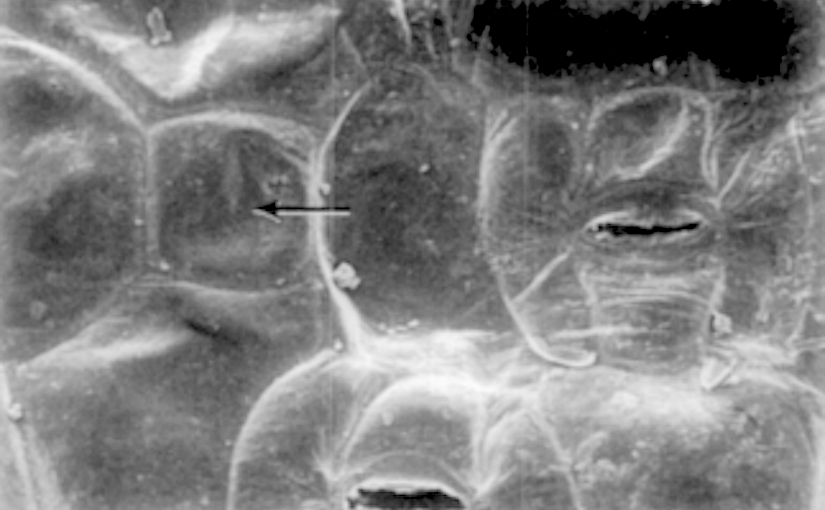
FIG. 4. A mature area of a Tradescantia leaf showing an arrested cell (arrow) and a mature, ablated stoma.


Stomatal patterning in Tradescantia: an evaluation of the cell lineage theory.

by Croxdale J., Smith J., Yandell B., Johnson J. B. (1992)

Judith Croxdale, Joseph Smith, Brian Yandell, and J. Bradley Johnson

Brian_Yandell
Brian S. Yandell, University of Wisconsin, Madison, USA

Department of Botany, University of Wisconsin, Madison 53706.

in Dev. Biol. 149:158–167 –

http://www.stat.wisc.edu/~yandell/doc/1992/25.DevelBio.pdf

Abstract

The cell lineage theory, which explains stomatal patterning in monocot leaves as a consequence of orderly divisions, was studied in Tradescantia.

Data were collected to test the theory at three levels of organization: the individual stoma; stomata distributed in one dimension, in linear fashion along cell files; and stomata apportioned in two dimensions, across the length and breadth of the leaf.

In an attempt to watch the patterning process through regeneration, stomata in all visible stages of development were laser ablated. The results showed that the formation of stomatal initials was highly regular, and measurements of stomatal frequency and spacing showed that pattern was determined near the basal meristem when the stomatal initials arose.

Following the origin of initials, the pattern was not readjusted by division of epidermal cells. Stomatal initials were not committed when first present and a small percentage of them arrested. The arrested cells, unlike stomata, were consistently positioned in cell files midway between a developed pair of stomata.

At the one-dimensional level of pattern, stomata in longitudinal files were separated by a variable number of epidermal cells and the frequency of these separations was not random.

The sequential spacing of stomata also was not random, and stomata separated by single epidermal cells were grouped into more short and long series than expected by chance.

The stomatal pattern across the width of the leaf resulted from cell files free of stomata which alternated with cell files containing stomata, but not with a recurring periodicity.

Files lacking stomata were found only over longitudinal vascular bundles. Laser ablations of developing stomata did not disrupt the pattern in nearby cells or result in stomatal regeneration.

We conclude that the cell lineage theory explains pattern as an individual stomatal initial arises from its immediate precursor and satisfactorily accounts for the minimum spacing of stomata in a cell file, i.e., stoma-epidermal cell-stoma.

However, the theory does not explain the collective stomatal pattern along the cell files, at the one-dimensional level of patterning. Nor does the theory account for the for the two-dimensional distribution of stomata in which regions devoid of stomata alternate with regions enriched with stomata, but not in a highly regular nor haphazard manner.

We suggest that the grouping of epidermal cells and stomata separated by single epidermal cells in cell files may result from cell lineages at a specific position in the cell cycle as they traverse the zone where stomatal initials form


Screen Shot 2017-09-15 at 14.25.20

Linear aggregations of stomata and epidermal cells


Linear aggregations of stomata and epidermal cells in Tradescantia leaves evidence for their group patterning as a function of the cell cycle.

by Chin J., Wan Y., Smith J., Croxdale J. (1995)

Department of Botany, University of Wisconsin, Madison 53706.

in Dev Biol 168:39–46 – DOI: 10.1006/dbio.1995.1059

https://www.ncbi.nlm.nih.gov/pubmed/7883077

Abstract

We tested Charlton’s hypothesis (1990) that stomata are present and patterned in linear cell aggregations using the monocot Tradescantia.

We examined the following features of the leaf epidermis in support of this theory: linear groups (strings) of stomatal complexes and of epidermal cells were sought in immature and mature regions of entire leaves; the lengths (in cell number) and incidences (numerical occurrence) of both string types were determined; the uniformity and progression of stomatal differentiation within strings were studied; physical characteristics of differentiating strings within cell files were measured.

Undifferentiated epidermal cells from the leaf base were stained with DAPI to reveal precursors of stomatal strings immediately proximal to the stomatal initial region.

The results indicated that the Tradescantia epidermis in the leaf blade consists of linear groups of stomata and epidermal cells, which did not change in cell number nor incidence during development.

The incidence of stomata by length was nonrandom. Although incidence decreased with string length, the decline was not linear nor exponential. Stomatal strings show cell cycle synchrony in DAPI staining of stomatal precursors and synchrony of stomatal differentiation within a string.

The irregularity in the length of the stomatal development region, and each differentiation stage in it, by cell file was consistent with the variation in string length and unity in string development.

The evidence supports Charlton’s hypothesis that cells are patterned based on their position in the cell cycle and that linear groups of stomata reflect cell lineages, which maintain a degree of cell cycle synchrony.