
In Vitro Propagation, Genetic Assessment, and Medium-Term Conservation of the Coastal Endangered Species Tetraclinis articulata (Vahl) Masters (Cupressaceae) from Adult Trees

 ,
,
Abstract
:1. Introduction
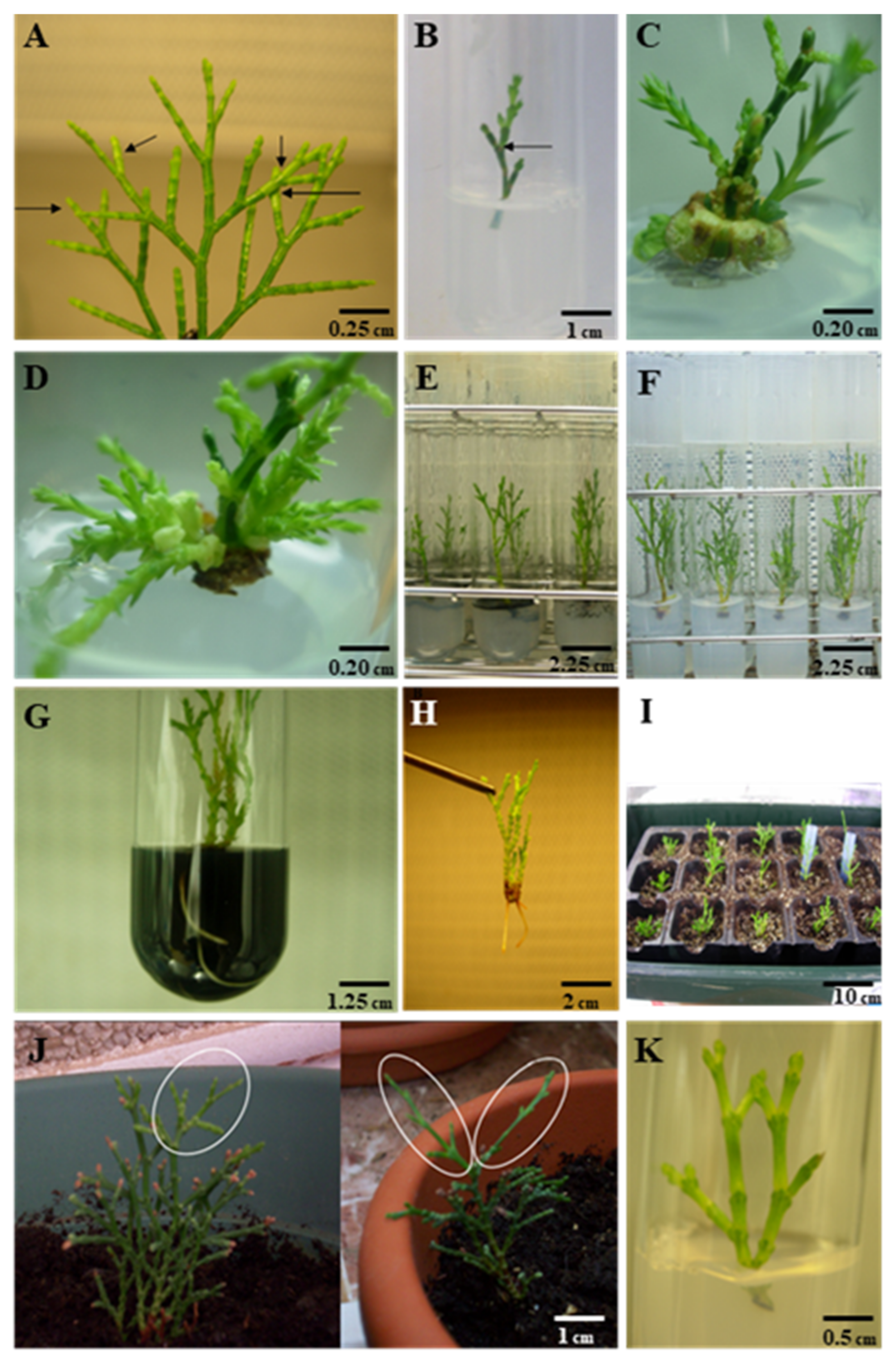
2. Results
2.1. Sterilization of the Plant Material and Culture Initiation
2.2. Growth Regulator Effects on Multiplication
2.3. Double-Phase Culture System (DPS) Effects on Shoot Elongation
2.4. Auxin Effects on Root Development
2.5. Culture Media Effects on Root Development
2.6. Extraction of Genomic DNA and RAPD Analysis
2.7. Acclimatization
2.8. Medium Term Storage of In Vitro Explants
3. Discussion
4. Materials and Methods
4.1. Sterilization of the Plant Material and Culture Initiation
4.2. Growth Regulator Effects on Multiplication
4.3. Double-Phase Culture System Effects on Shoot Elongation
4.4. Auxin Effects on Root Development
- Experiment 1.
- Explants 2–2.5 cm in length were transferred to SH basal medium supplemented with 30 g L−1 sucrose, 6.6 g L−1 agar, and auxins as follows: 0 (control) and 0.5 IBA, 0.5 NAA, and 0.5 g L−1 IBA plus 0.25 g L−1 NAA. A total amount of 16 explants was studied per replicate.
- Experiment 2.
- An auxin pulse treatment was performed on explants 2–2.5 cm in length taken from previously cultured plantlets on SH medium. The pulse treatment consisted of immersion of explants under dark conditions in liquid medium composed by SH salts, vitamins, and 250 mg of 2-(N-morpholino)ethanesulfonic acid (MES) as buffering agent to keep the culture pH between 5.75 and 5.8. Three concentrations (25, 50, and 100 mg L−1) of Indole Acetic Acid (IAA) at three immersion times (1, 2, and 4 h) were assayed. IAA was filter-sterilized and added to the previously described formulation after autoclaving. A sample without IAA was used as the control. After the treatment, explants were transferred to SH basal medium supplemented with 30 g L−1 sucrose, 6.6 g L−1 agar, and 0.5 g L−1 AC. Twenty-four explants were used per treatment and the rooting percentage was measured after 4, 6, and 8 weeks of culture.
4.5. Culture Media Effects on Root Development
4.6. Extraction of Genomic DNA and RAPD Analysis
4.7. Acclimatization
4.8. Medium-Term Storage of In Vitro Explants
4.9. Experimental Design and Data Analyses
5. Conclusions
Author Contributions
Funding
Institutional Review Board Statement
Informed Consent Statement
Data Availability Statement
Acknowledgments
Conflicts of Interest
References
- Quézel, P. Biogéographie et Écologie des Coniféres sur le Pourtour Méditerranéen in Actualités d’Écologie Forestière; Gauthier-Villars: Paris, France, 1980; ISBN 13-978-2842994518. [Google Scholar]
- Esteve Selma, M.A.; Montoya Bernabéu, P.; Moya Pérez, J.M.; Miñano Martínez, J.; Hernández García, I.; Carrión García, J.S.; Charco García, J.; Fernández Jiménez, S.; Munuera Giner, M.; Ochando Tomás, J. Tetraclinis articulata: Biogéographie, Écologie, Menaces et Conservation; Direction Générale du Milieu Naturel: Murcia, Spain, 2017; ISBN 978-84-09-08267-4. [Google Scholar]
- Elaieb, M.T.; Khouaja, A.; Khouja, M.L.; Valette, J.; Volle, G.; Candelier, K. Comparative Study of Local Tunisian Woods Properties and the Respective Qualities of Their Charcoals Produced by a New Industrial Eco-Friendly Carbonization Process. Waste Biomass Valorization 2018, 9, 1199–1211. [Google Scholar] [CrossRef]
- El Alami, S.; El Mouridi, M.; Laurent, T.; Calchéra, G.; Famiri, A.; Hakam, A.; Kabouchi, B.; Gril, J. Fracture energy of wood and root burl wood of Thuya (Tetraclinis articulata (Vahl) Masters). J. Trop. For. Sci. 2013, 25, 166–174. [Google Scholar]
- Fidah, A.; Salhi, N.; Janah, T.; Rahouti, M.; Kabouchi, B.; El Alami, A.; Ziani, M.; Famiri, A. Comparative natural durability of four Mediterranean softwoods against wood decay fungi. J. Indian Acad. Wood Sci. 2016, 13, 132–137. [Google Scholar] [CrossRef]
- Osete-Cortina, L.; Doménech-Carbó, M.T. Analytical characterization of diterpenoid resins present in pictorial varnishes using pyrolysis–gas chromatography–mass spectrometry with on line trimethylsilylation. J. Chromatogr. A 2005, 1065, 265–278. [Google Scholar] [CrossRef] [PubMed]
- Azémard, C.; Ménager, M.; Vieillescazes, C. On the tracks of sandarac, review and chemical analysis. Environ. Sci. Pollut. Res. 2017, 24, 27746–27754. [Google Scholar] [CrossRef]
- Jamila, F.; Mostafa, E. Ethnobotanical survey of medicinal plants used by people in Oriental Morocco to manage various ailments. J. Ethnopharmacol. 2014, 154, 76–87. [Google Scholar] [CrossRef] [PubMed]
- Bouyahya, A.; El Omari, N.; Elmenyiy, N.; Guaouguaou, F.-E.; Balahbib, A.; Belmehdi, O.; Salhi, N.; Imtara, H.; Mrabti, H.N.; El-Shazly, M.; et al. Moroccan antidiabetic medicinal plants: Ethnobotanical studies, phytochemical bioactive compounds, preclinical investigations, toxicological validations and clinical evidences; challenges, guidance and perspectives for future management of diabetes worldwide. Trends Food Sci. Technol. 2021, 115, 147–254. [Google Scholar] [CrossRef]
- Senouci, F.; Ababou, A.; Chouieb, M. Ethnobotanical survey of the medicinal plants used in the southern Mediterranean. Case study: The region of Bissa (Northeastern Dahra mountains, Algeria). Pharmacogn. J. 2019, 11, 647–659. [Google Scholar] [CrossRef] [Green Version]
- Jlizi, S.; Zardi-Bergaoui, A.; Znati, M.; Flamini, G.; Ascrizzi, R.; Ben Jannet, H. Chemical composition and biological evaluation of the resin from Tetraclinis articulata (Vahl.) Masters: A promising source of bioactive secondary metabolites. Ind. Crop. Prod. 2018, 124, 74–83. [Google Scholar] [CrossRef]
- Jlizi, S.; Lahmar, A.; Zardi-Bergaoui, A.; Ascrizzi, R.; Flamini, G.; Harrath, A.H.; Chekir-Ghedira, L.; Ben Jannet, H. Chemical composition and cytotoxic activity of the fractionated trunk bark essential oil from Tetraclinis articulata (Vahl) Mast. growing in Tunisia. Molecules 2021, 26, 1110. [Google Scholar] [CrossRef]
- Nicolás, M.J.; Esteve Selma, M.A.; Palazón, J.A.; López-Hernández, J.J. Modelo sobre las preferencias de hábitat a escala local de Tetraclinis articulata (Vahl) Masters en una población de su área de distribución. An. Biol. 2004, 26, 157–167. Available online: https://revistas.um.es/analesbio/article/view/30571 (accessed on 20 November 2021).
- Thomas, P. Tetraclinis articulata. The IUCN Red List of Threatened Species 2017, e.T30318A95804470. Available online: https://www.iucnredlist.org/es/species/30318/95804470 (accessed on 9 January 2022).
- Bañares, Á.; Blanca, G.; Güemes, J.; Moreno, J.C.; Ortiz, S. Atlas y Libro Rojo de la Flora Vascular Amenazada de España; Dirección General de Medio Natural y Política Forestal (Ministerio de MedioAmbiente, y Medio Rural y Marino)-Sociedad Española de Biología de la Conservación de Plantas: Madrid, Spain, 2011; ISBN 978-84-8014-795-8. [Google Scholar]
- Navarro Cerrillo, R.M.; del Campo, A.D.; Cortina, J. Factores que afectan al éxito de una repoblación y su relación con la calidad de la planta. In Calidad de Planta Forestal para la Restauración en Ambientes Mediterráneos. Estado Actual de Conocimientos; Cortina, J., Peñuelas, J.L., Puértolas, J., Vilagrosa, A., Savé, R., Eds.; Organismo Autónomo Parques Nacionales. Ministerio de Medio Ambiente: Madrid, Spain, 2006; ISBN 978-84-8014-670-8. [Google Scholar]
- Martínez-Sánchez, J.J.; Franco Leemhuis, J.A.; Vicente Colomer, M.J.; Muñoz, M.; Bañón Arias, S.; Conesa Gallego, E.; Fernández Hernández, J.A.; Valdés Illán, R.; Ochoa Rego, J.; Miralles Crespo, J.; et al. Especies Silvestres Mediterráneas con Valor Ornamental. Selección, Producción Viverística y Utilización en Jardinería; Serie Técnica No. 7; Servicio de Protección y Conservación de la Naturaleza. Dirección General de Patrimonio Natural y Biodiversidad. Consejería de Agricultura y Agua, Región de Murcia: Murcia, Spain, 2008; ISBN 978-84-691-8182-9. [Google Scholar]
- Bhojwani, S.S.; Dantu, P.K. Plant Tissue Culture: An Introductory Text; Springer: New Delhi, India, 2013; ISBN 9788132210269. [Google Scholar]
- Karimi, H.R.; Janghorban, K.; Raqamy, M.; Farahmand, H. In vitro propagation of some old Persian cypress accessions (Cupressus sempervirens L.) by embryo culture. Physiol. Mol. Biol. Plants 2018, 24, 1285–1294. [Google Scholar] [CrossRef]
- Ahn, C.H.; Heo, K.; Park, H.S.; Choi, Y.E. In vitro propagation and cryopreservation of Thuja koraiensis Nakai via somatic embryogenesis. In Vitro Cell. Dev. Biol. Plant 2019, 55, 605–614. [Google Scholar] [CrossRef]
- Von Aderkas, P.; Bonga, J.M. Influencing micropropagation and somatic embryogenesis in mature trees by manipulation of phase change, stress and culture environment. Tree Physiol. 2000, 20, 921–928. [Google Scholar] [CrossRef] [PubMed] [Green Version]
- Castro, M.R.; Belo, A.F.; Afonso, A.; Zavattieri, M.A. Micropropagation of Juniperus navicularis, an endemic and rare species from Portugal SW coast. Plant Growth Regul. 2011, 65, 223–230. [Google Scholar] [CrossRef] [Green Version]
- Loureiro, J.; Capelo, A.; Brito, G.; Rodriguez, E.; Silva, S.; Pinto, G.; Santos, C. Micropropagation of Juniperus phoenicea from adult plant explants and analysis of ploidy stability using flow cytometry. Biol. Plant. 2007, 51, 7–14. [Google Scholar] [CrossRef]
- Wang, Y.; Yao, R. Increased endogenous gibberellin level inhibits root growth of Pinus massoniana Lamb. plantlets during long-term subculture. In Vitro Cell. Dev. Biol. Plant 2020, 56, 470–479. [Google Scholar] [CrossRef]
- De Diego, N.; Montalbán, I.A.; Fernández de Larrinoa, E.; Moncaleán, P. In vitro regeneration of Pinus pinaster adult trees. Can. J. For. Res. 2008, 38, 2607–2615. [Google Scholar] [CrossRef]
- Morte, M.A.; Honrubia, M.; Piqueras, A. Micropropagation of Tetraclinis articulata (Vahl) Masters (Cupressaceae). Plant Cell Tissue Organ Cult. (PCTOC) 1992, 28, 231–233. [Google Scholar] [CrossRef]
- Momeni, M.; Ganji-Moghadam, E.; Kazemzadeh-Beneh, H.; Asgharzadeh, A. Direct Organogenesis from Shoot Tip Explants of Juniperus Polycarpos L.: Optimizing Basal Media and Plant Growth Regulators on Proliferation and Root Formation. Plant Cell Biotechnol. Mol. Biol. 2018, 19, 40–50. Available online: https://www.ikprress.org/index.php/PCBMB/article/view/1250 (accessed on 22 November 2021).
- Morte, M.; Diaz, G.; Honrubia, M. Effect of arbuscular mycorrhizal inoculation on micropropagated Tetraclinis articulata growth and survival. In Biotechnology in Agriculture and Forestry. Trees IV; Bajaj, Y.P.S., Ed.; Springer: Verlag, Germany, 1996; pp. 407–423. ISBN 978-3-642-59609-4. [Google Scholar]
- Benson, E. Plant Conservation Biotechnology; CRC Press: London, UK, 2014. [Google Scholar] [CrossRef]
- Weckx, S.; Inzé, D.; Maene, L. Tissue culture of oil palm: Finding the balance between mass propagation and somaclonal variation. Front. Plant Sci. 2019, 10, 722. [Google Scholar] [CrossRef] [Green Version]
- Komakech, R.; Kim, Y.-G.; Kim, W.J.; Omujal, F.; Yang, S.; Moon, B.C.; Okello, D.; Rahmat, E.; Kyeyune, G.N.; Matsabisa, M.G.; et al. A Micropropagation Protocol for the Endangered Medicinal Tree Prunus africana (Hook f.) Kalkman: Genetic Fidelity and Physiological Parameter Assessment. Front. Plant Sci. 2020, 11, 548003. [Google Scholar] [CrossRef] [PubMed]
- Serrano-Martinez, F.; Casas, J.L. Cryopreservation of Tetraclinis articulata (vahl.) Masters. CryoLetters 2011, 32, 248–255. [Google Scholar] [PubMed]
- Reed, B.M.; Chang, Y. Medium and long-term storage of in vitro cultures of temperate fruit and nut crops. In Conservation of Plant Genetic Resources In Vitro; Razdan, M.K., Cocking, E.C., Eds.; Science Publishers Inc.: London, UK, 1997; ISBN 978-1578080557. [Google Scholar]
- Reed, B.M.; Emgelmann, F.; Dulloo, M.E.; Engerls, J.M.M. Technical Guidance for the Management of Field and In Vitro Germplasm Collections. Handbooks for Genebanks No. 7; International Plant Genetic Resources Institute: Rome, Italy, 2018; 106p, Available online: https://www.bioversityinternational.org/e-library/publications/detail/technical-guidelines-for-the-management-of-field-and-in-vitro-germplasm-collections/ (accessed on 22 November 2021).
- Arbeloa, A.; Marín, J.A.; Andreu, P.; García, E.; Lorente, P. In vitro conservation of fruit trees by slow growth storage. Acta Hortic. 2017, 1156, 101–106. [Google Scholar] [CrossRef] [Green Version]
- Juan-Vicedo, J.; Ramírez-Luna, J.E.; Piqueras, A.; Casas, J.L. Micropropagation and cryopreservation by vitrification of the Spanish endemic medicinal plant Sideritis leucantha Cav. subsp. leucantha (Lamiaceae). In Vitro Cell. Dev. Biol. Plant 2021, 57, 1057–1065. [Google Scholar] [CrossRef]
- Ghareb, H.; Ibrahim, S.D.; Hegazi, G.E.M. Micropropagtion and DNA barcoding of the endangered endemic Phlomis aurea plant of Saint Katherine. Plant Omics 2020, 13, 65–77. [Google Scholar] [CrossRef]
- Phillips, G.C.; Garda, M. Plant tissue culture media and practices: An overview. In Vitro Cell. Dev. Biol. Plant 2019, 55, 242–257. [Google Scholar] [CrossRef]
- Juan-Vicedo, J.; Pavlov, A.; Ríos, S.; Casas, J.L. In vitro culture and micropropagation of the Baetic-Moroccan endemic plant Lapiedra martinezii Lag. (Amaryllidaceae). In Vitro Cell. Dev. Biol. Plant 2019, 55, 725–732. [Google Scholar] [CrossRef]
- Juan-Vicedo, J.; Pavlov, A.; Ríos, S.; Casas, J.L. Micropropagation of five endemic, rare and/or endangered Narcissus species from the Iberian Peninsula (Spain and Portugal). Acta Biol. Crac. Ser. Bot. 2021, 63, 55–61. [Google Scholar] [CrossRef]
- Thammasiri, K.; Jitsopakul, N.; Prasongsom, S. Micropropagation of some orchids and the use of cryopreservation. In Orchids Phytochemistry, Biology and Horticulture; Reference Series in Phytochemistry; Merillon, J.M., Kodja, H., Eds.; Springer: Cham, Switzerland, 2020; pp. 1–36. [Google Scholar] [CrossRef]
- Hazubska-Przybył, T.; Przybył, H. Propagation of Juniper species by plant tissue culture: A mini-review. Forests 2019, 10, 1028. [Google Scholar] [CrossRef] [Green Version]
- Kumar, N.; Reddy, M.P. In vitro plant propagation: A review. J. For. Sci. 2011, 27, 61–72. [Google Scholar]
- Gaj, M.D. Factors influencing somatic embryogenesis induction and plant regeneration with particular reference to Arabidopsis thaliana (L.) Heynh. Plant Growth Regul. 2004, 43, 27–47. [Google Scholar] [CrossRef]
- Mostafa, H.H.A.; Wang, H.; Song, J.; Li, X. Effects of genotypes and explants on garlic callus production and endogenous hormones. Sci. Rep. 2020, 10, 4867. [Google Scholar] [CrossRef] [Green Version]
- Zhang, H.; Horgan, K.J.; Reynolds, P.H.S.; Jameson, P.E. 6-Benzyladenine metabolism during reinvigoration of mature Pinus radiata buds in vitro. Tree Physiol. 2010, 30, 514–526. [Google Scholar] [CrossRef] [Green Version]
- Galán-Ávila, A.; García-Fortea, E.; Prohens, J.; Herraiz, F.J. Development of a direct in vitro plant regeneration protocol from Cannabis sativa L. seedling explants: Developmental morphology of shoot regeneration and ploidy level of regenerated plants. Front. Plant Sci. 2020, 11, 645. [Google Scholar] [CrossRef] [PubMed]
- Vieira, L.M.; Rocha, D.I.; Taquetti, M.F.; Da Silva, L.C.; De Campos, J.M.S.; Viccini, L.F.; Otoni, W.C. In vitro plant regeneration of Passiflora setacea D.C. (Passifloraceae): The influence of explant type, growth regulators, and incubation conditions. In Vitro Cell. Dev. Biol. Plant 2014, 50, 738–745. [Google Scholar] [CrossRef]
- Pullman, G.S.; Skryabina, A. Liquid medium and liquid overlays improve embryogenic tissue initiation in conifers. Plant Cell Rep. 2007, 26, 873–887. [Google Scholar] [CrossRef]
- Gross, L.M. Micropropagation of Douglas Fir (Pseudotsuga menziesii) in a Double-Phase Culture Medium. Ph.D. Thesis, Oregon State University, Corvallis, OR, USA, 2002. [Google Scholar]
- Ćosić, T.; Motyka, V.; Savić, J.; Raspor, M.; Marković, M.; Dobrev, P.I.; Ninković, S. Sucrose interferes with endogenous cytokinin homeostasis and expression of organogenesis-related genes during de novo shoot organogenesis in kohlrabi. Sci. Rep. 2021, 11, 1–16. [Google Scholar] [CrossRef]
- Aslam, F.; Naz, S.; Tariq, A.; Ilyas, S.; Shahzadi, K. Rapid multiplication of ornamental bulbous plants. Pak. J. Bot. 2013, 45, 2051–2055. [Google Scholar]
- Yaseen, M.; Ahmad, T.; Sablok, G.; Standardi, A.; Hafiz, I.A. Review: Role of carbon sources for in vitro plant growth and development. Mol. Biol. Rep. 2013, 40, 2837–2849. [Google Scholar] [CrossRef]
- Maene, L.; Debergh, P. Liquid medium additions to established tissue cultures to improve elongation and rooting in vivo. Plant Cell Tissue Organ Cult. (PCTOC) 1985, 5, 23–33. [Google Scholar] [CrossRef]
- Piqueras, A.; Han, B.; Van Huylenbroeck, J.; Debergh, P. Effect of different environmental conditions in vitro on sucrose metabolism and antioxidant enzymatic activities in cultured shoots of Nicotiana tabacum L. Plant Growth Regul. 1998, 25, 5–10. [Google Scholar] [CrossRef]
- Cano-Castillo, M.; Serrano-Martínez, F.; Casas, J.L. In vitro propagation of Astragalus nitidiflorus (Leguminosae), an endemic and endangered species from South-East of Spain. Acta Hortic. 2009, 812, 545–550. [Google Scholar] [CrossRef]
- Khamushi, M.; Dehestani-Ardakani, M.; Zarei, A.; Aliabad, K.K. An efficient protocol for micropropagation of old cypress of Abarkuh (Cupressus sempervirens var. horizontalis [Mill.]) under in vitro condition. Plant Cell Tissue Organ Cult. (PCTOC) 2019, 138, 597–601. [Google Scholar] [CrossRef]
- Patel, A.K.; Lodha, D.; Shekhawat, N.S. An improved micropropagation protocol for the ex situ conservation of Mitragyna parvifolia (Roxb.) Korth. (Rubiaceae): An endangered tree of pharmaceutical importance. In Vitro Cell. Dev. Biol. Plant 2020, 56, 817–826. [Google Scholar] [CrossRef]
- Bairu, M.W.; Aremu, A.O.; Van Staden, J. Somaclonal variation in plants: Causes and detection methods. Plant Growth Regul. 2011, 63, 147–173. [Google Scholar] [CrossRef]
- Panis, B.; Nagel, M.; Van den Houwe, I. Challenges and prospects for the conservation of crop genetic resources in field genebanks, in in vitro collections and/or in liquid nitrogen. Plants 2020, 9, 1634. [Google Scholar] [CrossRef] [PubMed]
- Żabicki, P.; Sliwinska, E.; Mitka, J.; Sutkowska, A.; Tuleja, M.; Migdałek, G.; Żabicka, J.; Słomka, A.; Kwiatkowska, M.; Kuta, E. Does somaclonal variation play advantageous role in conservation practice of endangered species?: Comprehensive genetic studies of in vitro propagated plantlets of Viola stagnina Kit. (Violaceae). Plant Cell, Tissue Organ Cult. (PCTOC) 2019, 136, 339–352. [Google Scholar] [CrossRef]
- Ranghoo-Sanmukhiya, V.M. Somaclonal variation and methods used for its detection. In Propagation and Genetic Manipulation of Plants; Siddique, I., Ed.; Springer: Singapore, 2021; pp. 1–18. [Google Scholar] [CrossRef]
- Oliya, B.K.; Chand, K.; Thakuri, L.S.; Baniya, M.K.; Sah, A.K.; Pant, B. Assessment of genetic stability of micropropagated plants of Rhynchostylis retusa (L.) using RAPD markers. Sci. Hortic. 2021, 281, 110008. [Google Scholar] [CrossRef]
- Novikova, T.I.; Asbaganov, S.V.; Ambros, E.V.; Zaytseva, Y.G. TDZ-induced axillary shoot proliferation of Rhododendron mucronulatum Turcz and assessment of clonal fidelity using DNA-based markers and flow cytometry. In Vitro Cell. Dev. Biol. Plant 2020, 56, 307–317. [Google Scholar] [CrossRef]
- Swain, D.; Pushpalatha, G.; Das, P. In vitro micro-propagation of Typhonium flagelliforme (Lodd.) Blume and its genetic fidelity using ISSR and RAPD markers. J. Pharmacogn. Phytochem. 2020, 9, 3484–3488. [Google Scholar]
- Rozas, V.; García-Cervigón, A.I.; García-Hidalgo, M.; Rodríguez-García, E.; Olano, J.M. Living on the edge: Legacy of water availability on Tetraclinis articulata secondary growth under semiarid conditions in Morocco. Dendrochronologia 2021, 68, 125853. [Google Scholar] [CrossRef]
- Oliveras, I.; Martínez-Vilalta, J.; Jimenez-Ortiz, T.; Lledó, M.J.; Escarré, A.; Pinol, J. Hydraulic properties of Pinus halepensis, Pinus pinea and Tetraclinis articulata in a dune ecosystem of Eastern Spain. Plant Ecol. 2003, 169, 131–141. [Google Scholar] [CrossRef]
- Pospíšilová, J.; Tichá, I.; Kadleček, P.; Haisel, D.; Plzáková, S. Acclimatization of micropropagated plants to ex vitro conditions. Biol. Plant. 1999, 42, 481–497. [Google Scholar] [CrossRef]
- Hazarika, B.N. Acclimatization of tissue-cultured plants. Curr. Sci. 2003, 85, 1704–1712. Available online: https://www.jstor.org/stable/24109975 (accessed on 25 November 2021).
- Alzubi, H.; Yepes, L.M.; Fuchs, M. In vitro storage of micropropagated grapevine rootstocks at low temperature. In Vitro Cell. Dev. Biol. Plant 2019, 55, 334–341. [Google Scholar] [CrossRef]
- Ballester, A.; Janeiro, L.V.; Vieitez, A.M. Cold storage of shoot cultures and alginate encapsulation of shoot tips of Camellia japonica L. and Camellia reticulata Lindley. Sci. Hortic. 1997, 71, 67–78. [Google Scholar] [CrossRef]
- Romano, A.; Martins-Loução, M.A. In vitro cold storage of cork oak shoot cultures. Plant Cell Tissue Organ Cult. (PCTOC) 1999, 59, 155–157. [Google Scholar] [CrossRef]
- Ozudogru, E.A.; Kirdok, E.; Kaya, E.; Capuana, M.; Benelli, C. In vitro conservation of red-wood (Sequoia sempervirens) by slow growth storage and cryopreservation. Acta Hortic. 2012, 961, 291–296. [Google Scholar] [CrossRef]
- Schenk, R.U.; Hildebrandt, A.C. Medium and techniques for induction and growth of monocotyledonous and dicotyledonous plant cell cultures. Can. J. Bot. 1972, 50, 199–204. [Google Scholar] [CrossRef]
- Knop, W. Quantitative untersuchungen über die ernahrungsprocesse der pflanzen. Landwirtsch. Vers.-Station. 1865, 7, 93–107. [Google Scholar]


| Treatment (mg L−1) | Length Increment (cm) | Explants with Basal Shoots (%) | Basal Shoots per Explant (nº) | |||
|---|---|---|---|---|---|---|
| 6 Weeks | 12 Weeks | 6 Weeks | 12 Weeks | 6 Weeks | 12 Weeks | |
| BAP | ||||||
| 0 | 1.60 ± 0.21 a | 2.86 ± 0.26 a | 0.00 ± 00.0 c | 0.00 ± 0.00 e | 0.00 ± 0.00 c | 0.00 ± 0.00 e |
| 0.5 | 0.98 ± 0.08 bcd | 2.30 ± 0.22 bc | 13.81 ± 4.27 ab | 53.49 ± 2.06 bc | 0.32 ± 0.10 ab | 1.17 ± 0.12 d |
| 1.0 | 0.76 ± 0.06 cde | 1.44 ± 0.10 efg | 20.21 ± 4.20 a | 79.85 ± 5.32 a | 0.47 ± 0.19 a | 2.60 ± 0.43 a |
| 2.0 | 0.51 ± 0.05 def | 1.26 ± 0.04 g | 10.70 ± 5.58 abc | 68.89 ± 4.44 a | 0.24 ± 0.12 abc | 1.96 ± 0.22 b |
| 4.0 | 0.47 ± 0.04 ef | 1.32 ± 0.09 fg | 4.31 ± 2.16 cb | 65.42 ± 4.97 ab | 0.06 ± 0.04 bc | 1.85 ± 0.05 b |
| KIN | ||||||
| 0.5 | 1.14 ± 0.04 b | 2.71 ± 0.02 ab | 0.00 ± 0.00 c | 4.17 ± 2.08 e | 0.00 ± 0.00 c | 0.04 ± 0.02 e |
| 1.0 | 1.00 ± 0.11 bc | 2.18 ± 0.17 cd | 0.00 ± 0.00 c | 4.44 ± 2.22 e | 0.00 ± 0.00 c | 0.09 ± 0.06 e |
| 2.0 | 0.69 ± 0.13 ef | 1.89 ± 0.09 cde | 8.61 ± 1.94 bc | 24.4 ± 8.01 d | 0.22 ± 0.04 abc | 0.69 ± 0.20 cd |
| 4.0 | 0.41 ± 0.04 f | 1.82 ± 0.19 de | 11.43 ± 2.40 ab | 46.51 ± 2.06 c | 0.23 ± 0.06 abc | 1.19 ± 0.14 d |
| TDZ | ||||||
| 0.025 | 1.84 ± 0.09 a | 2.89 ± 0.17 a | 10.41 ± 2.08 abc | 29.72 ± 3.92 d | 0.19 ± 0.06 bc | 0.74 ± 0.07 d |
| 0.05 | 0.95 ± 0.08 bcd | 1.75 ± 0.17 def | 6.25 ± 3.61 bc | 27.08 ± 4.17 d | 0.08 ± 0.06 bc | 0.48 ± 0.11 de |
| 0.1 | 0.51 ± 0.11 ef | 1.12 ± 0.12 gh | 4.17 ± 4.17 bc | 29.86 ± 9.19 d | 0.06 ± 0.06 bc | 0.32 ± 0.14 de |
| 0.25 | 0.57 ± 0.03 ef | 0.76 ± 0.06 h | 4.17 ± 4.17 bc | 6.25 ± 6.25 e | 0.06 ± 0.06 bc | 0.10 ± 0.10 e |
| Treatment (mg L−1) | Length Increment (cm) | Explants with Basal Shoots (%) | Basal Shoots per Explant (nº) | ||||
|---|---|---|---|---|---|---|---|
| 6 Weeks | 12 Weeks | 6 Weeks | 12 Weeks | 6 Weeks | 12 Weeks | ||
| BAP | NAA | ||||||
| 0 | 0 | 2.24 ± 0.20 a | 3.26 ± 0.13 a | 0.00 ± 0.00 c | 0.00 ± 0.00 d | 0.00 ± 0.00 d | 0.00 ± 0.00 e |
| 0.5 | 0.05 | 1.28 ± 0.10 b | 2.83 ± 0.20 ab | 58.33 ± 8.55 a | 84.29 ± 8.81 a | 1.36 ± 0.19 ab | 2.40 ± 0.31 ab |
| 1.0 | 0.05 | 0.67 ± 0.04 c | 1.28 ± 0.18 de | 59.68 ± 13.75 a | 77.38 ± 10.40 ab | 1.51 ± 0.27 a | 3.04 ± 0.25 a |
| 2.0 | 0.05 | 0.42 ± 0.06 bcde | 0.65 ± 0.07 ef | 27.78 ± 7.73 b | 75.56 ± 9.69 ab | 0.62 ± 0.14 c | 2.11 ± 0.09 bc |
| 4.0 | 0.05 | 0.19 ± 0.03 e | 0.38 ± 0.07 f | 38.61 ± 12.55 ab | 90.48 ± 9.52 a | 0.86 ± 0.27 bc | 2.58 ± 0.29 ab |
| KIN | NAA | ||||||
| 0.5 | 0.05 | 1.11 ± 0.04 b | 3.05 ± 0.29 a | 30.28 ± 12.08 ab | 46.11 ± 12.03 c | 0.54 ± 0.21 cd | 0.99 ± 0.29 d |
| 1.0 | 0.05 | 0.49 ± 0.02 cd | 2.17 ± 0.32 bc | 41.67 ± 4.16 ab | 56.55 ± 3.62 bc | 1.00 ± 0.19 ab | 1.60 ± 0.03 c |
| 2.0 | 0.05 | 0.41 ± 0.09 cde | 1.61 ± 0.37 cd | 31.75 ± 4.16 ab | 66.67 ± 6.30 abc | 0.75 ± 0.17 c | 2.00 ± 0.18 bc |
| 4.0 | 0.05 | 0.30 ± 0.03 de | 1.35 ± 0.16 d | 34.70 ± 6.10 ab | 73.50 ± 5.60 ab | 0.65 ± 0.08 c | 2.03 ± 0.11 bc |
| No. | Primer Code | Primer Sequences (5′-3′) |
|---|---|---|
| 16 | BAS16 | GCGATTTGCC |
| 21 | BAS21 | GAGCAGCGAA |
| 26 | BAS26 | CGGACGCATT |
| 27 | BAS27 | ACGCTGGTAG |
| 29 | BAS29 | CCACGCAACA |
Publisher’s Note: MDPI stays neutral with regard to jurisdictional claims in published maps and institutional affiliations. |
© 2022 by the authors. Licensee MDPI, Basel, Switzerland. This article is an open access article distributed under the terms and conditions of the Creative Commons Attribution (CC BY) license (https://creativecommons.org/licenses/by/4.0/).
Share and Cite
Juan-Vicedo, J.; Serrano-Martínez, F.; Cano-Castillo, M.; Casas, J.L. In Vitro Propagation, Genetic Assessment, and Medium-Term Conservation of the Coastal Endangered Species Tetraclinis articulata (Vahl) Masters (Cupressaceae) from Adult Trees. Plants 2022, 11, 187. https://doi.org/10.3390/plants11020187
Juan-Vicedo J, Serrano-Martínez F, Cano-Castillo M, Casas JL. In Vitro Propagation, Genetic Assessment, and Medium-Term Conservation of the Coastal Endangered Species Tetraclinis articulata (Vahl) Masters (Cupressaceae) from Adult Trees. Plants. 2022; 11(2):187. https://doi.org/10.3390/plants11020187
Chicago/Turabian StyleJuan-Vicedo, Jorge, Francisco Serrano-Martínez, Miriam Cano-Castillo, and José Luis Casas. 2022. "In Vitro Propagation, Genetic Assessment, and Medium-Term Conservation of the Coastal Endangered Species Tetraclinis articulata (Vahl) Masters (Cupressaceae) from Adult Trees" Plants 11, no. 2: 187. https://doi.org/10.3390/plants11020187