
Recommended
More Related Content
What's hot
What's hot (20)
Similar to Hemangioma
Similar to Hemangioma (20)
Recently uploaded
Recently uploaded (20)
Hemangioma
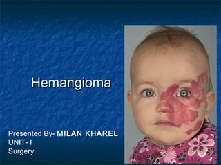
- 1. HemangiomaHemangioma Presented By- MILAN KHAREL UNIT- I Surgery
- 2. IntroductionIntroduction •• It is benign vascular endothelial tumour, common inIt is benign vascular endothelial tumour, common in girls (3 : 1).girls (3 : 1). •• It is commonly seen in skin and subcutaneous tissueIt is commonly seen in skin and subcutaneous tissue •• It grows rapidly in first year and 70 % involutes inIt grows rapidly in first year and 70 % involutes in 7 years.7 years. •• Early proliferative lesion is bright red, irregular; deepEarly proliferative lesion is bright red, irregular; deep lesion is bluish coloured. Involution causes colourlesion is bluish coloured. Involution causes colour fading, softness, shrinkage leaving crepe paper likefading, softness, shrinkage leaving crepe paper like area.area.
- 3. ClassificationClassification •• CapillaryCapillary –– Salmon patch (stork bite)Salmon patch (stork bite) –– Strawberry haemangiomaStrawberry haemangioma –– Port wine stainPort wine stain •• CavernousCavernous
- 4. clinical appearanceclinical appearance depends on its depth relative to the dermis anddepends on its depth relative to the dermis and the growth phase.the growth phase. Early proliferating lesionsEarly proliferating lesions are bright red,are bright red, irregularly surfaced,irregularly surfaced, papular lesions reminiscent of strawberries;papular lesions reminiscent of strawberries; deeper lesionsdeeper lesions may be blue or skin coloured.may be blue or skin coloured.
- 5. InvolutionInvolution typically begins with the fadingtypically begins with the fading of colour, leaving greyish areas, and theof colour, leaving greyish areas, and the lesion softens and shrinks at the samelesion softens and shrinks at the same time.time. When involution is complete, often allWhen involution is complete, often all that remains is the ‘crêpe paper’-texturedthat remains is the ‘crêpe paper’-textured area of skinarea of skin
- 6. The majority require no specific treatment. intervention during the early proliferative growth phase. Periorbital can cause 1. deprivational amblyopia or 2. Total obstruction of one visual field may result in permanent visual impairment. 3. astigmatism So, ophthalmologist should be involved immediately.
- 7. large haemangiomatalarge haemangiomata obstruct the nose, pose a serious risk.obstruct the nose, pose a serious risk. Rarely, skin ulceration leading to haemorrhage .Rarely, skin ulceration leading to haemorrhage . Blood-borneBlood-borne bacteria septicaemia and localised necrosis,bacteria septicaemia and localised necrosis, thrombocytopenia (Kasabach–Merrittthrombocytopenia (Kasabach–Merritt syndrome) andsyndrome) and large visceral or multiple lesions can cause congestivelarge visceral or multiple lesions can cause congestive heart failure secondary to shunting of blood.heart failure secondary to shunting of blood.
- 8. TREATMENTTREATMENT The management of most clinicalThe management of most clinical problems is primarily nonsurgical.problems is primarily nonsurgical. Systemic corticosteroids induce involution in upSystemic corticosteroids induce involution in up to 60%to 60% (2 mg kg-1 for 3 weeks, then a tapering dose).(2 mg kg-1 for 3 weeks, then a tapering dose). The primary role ofThe primary role of surgery is in the management of abnormallysurgery is in the management of abnormally textured or redundant skin left behind aftertextured or redundant skin left behind after involution.involution.
- 10. Salmon patchSalmon patch A haemangioma that presents as a pinkA haemangioma that presents as a pink macule, usually at themacule, usually at the nape of the neck, in 50% of infants. It isnape of the neck, in 50% of infants. It is caused by an area of persistentcaused by an area of persistent fetal dermal circulation and usuallyfetal dermal circulation and usually disappears within a year.disappears within a year.
- 12. Capillary haemangiomaCapillary haemangioma (strawberry)(strawberry) The commonest birthmark, commonly onThe commonest birthmark, commonly on the head and neck;the head and neck; 90% appear at birth and, as a consequence of90% appear at birth and, as a consequence of intravascular thrombosis, fibrosis and mast cellintravascular thrombosis, fibrosis and mast cell infiltration,infiltration, 10% resolve each subsequent year, with 70% resolved10% resolve each subsequent year, with 70% resolved by the age of 7 years.by the age of 7 years. White-skinned individualsWhite-skinned individuals girls are affected three times more than boys.girls are affected three times more than boys.
- 14. ‘‘Port-wine’ stainsPort-wine’ stains Equal gender distribution Birth prevalence is 0.3% 20 times less common than strawberry haemangiomata; “port-wine stains,” are present at birth as permanent, flat, pink-red cutaneous lesions
- 15. Defective maturation of cutaneous sympathetic innervation during embryogenesis, leading to localised intradermal capillary vasodilatation. At birth, they appear as flat, smooth, intensely purplestained areas, most frequently on the head and neck, often within the maxillary and mandibular dermatomes of the trigeminal nerve; with age their surfaces become more keratotic and nodular.
- 16. TreatmentTreatment • Allowed for spontaneous regression. • Otherwise by laser therapy – pulsed dye laser (diode laser). • CO2 snow therapy. • Sclerosant therapy. • Steroid - oral / intralesional / systemic. • Therapeutic embolisation
- 17. Port-wine stain (PWS) is associated with several syndromes:Port-wine stain (PWS) is associated with several syndromes: •• Sturge–Weber syndromeSturge–Weber syndrome. PWS affecting trigeminal dermatomes;. PWS affecting trigeminal dermatomes; associated with epilepsy and glaucoma (secondary to ipsilateral,associated with epilepsy and glaucoma (secondary to ipsilateral, leptomeningeal angiomatosis), cortical atrophy and visual fieldleptomeningeal angiomatosis), cortical atrophy and visual field defects.defects. •• Klippel–Trenaunay–Weber syndromeKlippel–Trenaunay–Weber syndrome. PWS on a limb with associated. PWS on a limb with associated bone and soft tissue hypertrophy and lateral varicosebone and soft tissue hypertrophy and lateral varicose veins when the lower limb is involved. It is calledveins when the lower limb is involved. It is called Parkes–Weber syndrome if there is an associated arteriovenousParkes–Weber syndrome if there is an associated arteriovenous malformation.malformation. •• Proteus syndromeProteus syndrome. PWS and regional gigantism in association. PWS and regional gigantism in association with lymphatic (lymphaticovenous) malformation. Hypertrophywith lymphatic (lymphaticovenous) malformation. Hypertrophy is always asymmetrical.is always asymmetrical.
- 18. Cavernous HaemangiomaCavernous Haemangioma • It is present at birth and consists of a multiple venous channels. • Its size increases gradually and may cause problems. • Sites: Head, neck, face, limbs, tongue, liver and other internal organs. • Large or multiple cavernous haemangiomas can cause congestive heart failure (hyperdynamic) due to shunting of large quantity of blood.
- 20. Clinical FeaturesClinical Features • It is smooth, soft, well-localised, warm, fluctuant, compressible, nonpulsatile swelling with bluish surface occurring in skin and subcutaneous tissue (often in mucosa like oral cavity) without any transillumination. • Compressibility and bluish surface is diagnostic.
- 21. TreatmentTreatment •• Sclerosant therapySclerosant therapy • Ligation of feeding artery and often at later stage excision is done once haemangioma shrinks. • Therapeutic embolisation. • If small and located in accessible area, excision is the initial therapy. • Laser ablation – diode pulsed laser is becoming popular because of good control of bleeding.